Metallographic Examination
Notice: Undefined index: background_image in /home/mtalabor/public_html/wp-content/plugins/elementor/includes/conditions.php on line 87
Notice: Trying to access array offset on value of type null in /home/mtalabor/public_html/wp-content/plugins/elementor/includes/conditions.php on line 90
Notice: Undefined index: background_image in /home/mtalabor/public_html/wp-content/plugins/elementor/includes/conditions.php on line 87
Notice: Trying to access array offset on value of type null in /home/mtalabor/public_html/wp-content/plugins/elementor/includes/conditions.php on line 90
Notice: Undefined index: background_image in /home/mtalabor/public_html/wp-content/plugins/elementor/includes/conditions.php on line 87
Notice: Trying to access array offset on value of type null in /home/mtalabor/public_html/wp-content/plugins/elementor/includes/conditions.php on line 90
Notice: Undefined index: background_image in /home/mtalabor/public_html/wp-content/plugins/elementor/includes/conditions.php on line 87
Notice: Trying to access array offset on value of type null in /home/mtalabor/public_html/wp-content/plugins/elementor/includes/conditions.php on line 90
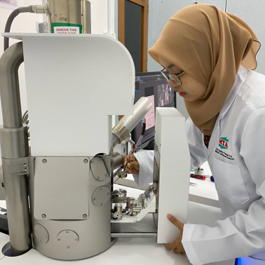
Microstructure and Elemental Analysis
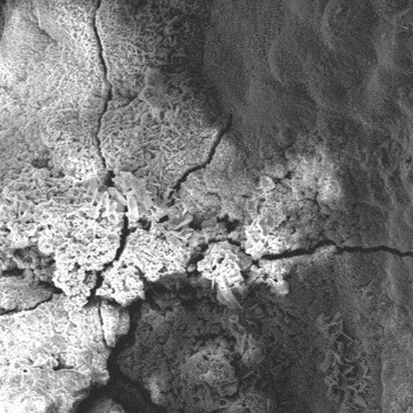
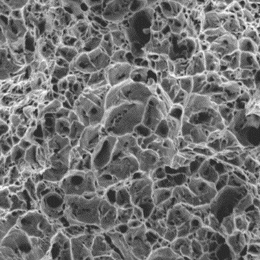
Scanning electron microscope is a type of microscope that uses a high-energy beam of electrons to illuminate a specimen and create a highly-magnified image over 100 000 times magnification and contain the information about the sample’s surface topography, composition and other properties such as
- Detect and analyze surface fracture.
- Examine surface contamination.
- Provide mapping and elemental analysis (EDX)
- Capable to Provide metallography analysis for nonconductive sample
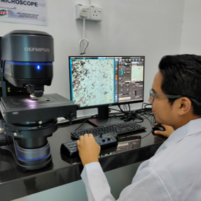
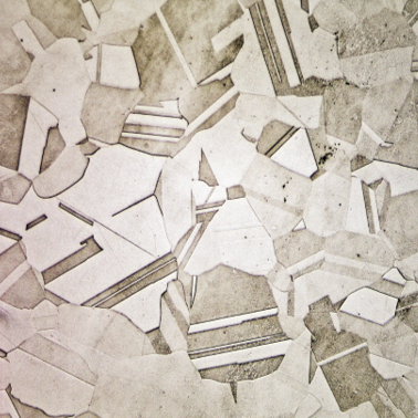
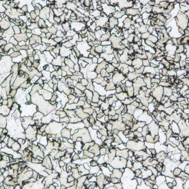
Micro & Macro Metallographic Examination
MTA Laboratory provide Imaging of topographic or microstructural features on polished and etched surfaces at magnifications of 140x up to 1400× Macro to Micro Versatility. By using 2 type of microscope we can accurately identify the phase analysis, grain intercept, grain count and measure in high quality image with High-Resolution Image at High Magnification. With the high end equipment with a single click we can Choose from 6 observation methods that can observe the sample through 180 degree.